Temporary Blindness

Please register or login

Welcome to ScubaBoard, the world's largest scuba diving community. Registration is not required to read the forums, but we encourage you to join. Joining has its benefits and enables you to participate in the discussions.

Benefits of registering include

  • Ability to post and comment on topics and discussions.
  • A Free photo gallery to share your dive photos with the world.
  • You can make this box go away

Joining is quick and easy. Log in or Register now!

I did the examination of tonometry / color photo of fundus / visual field on my eyes, the doctor said the results showed no problem. But I do not know which result represents my retinal examination was normal. (I did not say to Kelvin my retinal examination was normal), I think color photo of fundus for retinal examination, is that right?

Hi aviayu,

Tonometry measures the pressure of the fluid within the eye. It's used in the diagnosis of glaucoma.

Ocular fundus photography provides a crisp photo of the retina, retinal vasculature and head of the optic nerve/optic disc.

The retina also is often examined by dilating the eyes with drops and then observing the retinas witn an ophthalmoscope (a lighted magnifying device). Makes you look like an owl for awhile.

Regards,

DocVikingo
 
The reason for the questions about how the blindness progressed is that it may give clues to where a problem may be. The visual pathway goes from the eyes, then along optic nerves which divide, finally ending in the back of the brain. Problems in different areas can give different patterns of blindness.

Your pattern of first one eye and then the other becoming blind seems to make it more likely that the problem is with the eyes themselves or along the optic nerves immediately behind the eyes and before going deeper into the brain (esp. before the optic chiasm). If the problem was deeper in the brain, strange patterns like half-eye blindness occur. And if the problem was with the back of the brain where all the visual signals are gathered and processed, then it would be very likely that both eyes would be affected at the same time.

It is also important to realize that the problem might not be with the optic nerves themselves but with their supporting nutrient blood flows. The retinal artery was been mentioned by DDM but just as quickly noted that the blindness pattern of "curtain falling" is not typical of a disruption of blood flow there.

It is good that your eyes have been checked. However, it is important to be very assured that your retinas are ok. Standard ophthalmoscope examination might not be good enough; a special ultrasound examination may be appropriate. It seems likely that for both eyes to be affected, there is a common problem; e.g. congential to the eyes or a structure which supports both the eyes.
 
Last edited:
Happy to know “diving thus far has not been shown to be a problem for glaucoma patients”. Do you know any cases which was similar with my case(temporary blind). Even I have glaucoma, it seems to be a incipient/subclinical glaucoma or normal pressure/normal tension glaucoma, is it really a problem that affect my diving?

I have not heard of such a case, but that doesn't mean they don't exist.

The pieces cited in my post above appear to indicate that incipient/subclinical glaucoma and normal pressure glaucoma are not a particular worry as regards SCUBA, but you'll still want clearance from your ophthalmologist.

Regards,

DocVikingo

This is educational only and does not constitute or imply a doctor-patient relationship. It is not medical advice to you or any other individual and should not be construed as such.
 
It is good that your eyes have been checked. However, it is important to be very assured that your retinas are ok. Standard ophthalmoscope examination might not be good enough; a special ultrasound examination may be appropriate.

Hi cutlass,

Given that the interior of the globe can be fully and clearly seen, is "a special ultrasound examination" (high-resolution ultrasound imaging?) still considered more sensititive than ocular fundus photography? Does it add significant additional information? I guess it might give a better picture of deeper tissue structures.

Thanks,

Doc
 
If y'all will indulge me for a minute, I'd like to summarize what we have so far, along with the short version of some consultations:

A healthy 50 year old male with no significant past medical history makes a dive, breathing gas unknown but presumed to be air or nitrox. He reports no difficulty equalizing his ears or sinuses, and equalizes via the Toynbee (pinch and swallow) maneuver vs. the Valsalva maneuver. On descent he noticed decreased vision in his right eye but assumed it to be related to mask fogging and continued his descent. At 22 meters (72 FSW) he reported complete loss of vision in the right eye. He continued his descent, and at a depth of 26 meters (85 FSW) he reported a complete loss of vision.
He immediately made a controlled ascent with the assistance of the dive guide, and reported an improvement of vision at a depth of 7-8 meters (23-26 FSW) with complete resolution of symptoms upon reaching the surface. At the surface, it was noted that his eyes were red and the area around his eyes was swollen. There was a small amount of blood around the seal of the mask. The divemaster on the boat diagnosed mask squeeze, but the diver and his buddy are not sure, as they have seen severe mask squeeze and that diver's appearance was different. Exact description of the appearance of his eyes is lacking, and there are no photographs.
Complete exam by an ophthalmologist an unknown time after the dive reveals no abnormalities. Intraocular pressures are normal, and a fundoscopic exam of the retinas is unremarkable.

DocV and Cutlass, does this sound pretty accurate to y'all?

What we've already pretty much ruled out:
1. O2 toxicity.
2. Open angle glaucoma, narrow angle glaucoma, normal pressure glaucoma, and bad glaucomb-over.
3. Acute retinal detachment.

So far, I've spoken with two of our attendings, Dr. Jake Freiberger and Dr. Richard Moon; chief DAN medic Dan Nord; and CAPT Frank Butler, USN (RET) CAPT Butler is a well-known Navy diving medical officer with a specialty in ophthalmology. Here are the opinions:

Before hearing the results of the ophthalmology visit, Dr. Freiberger believed this was a mask squeeze, worrisome for possible glaucoma aggravation.

Dr. Moon believes it's a mask squeeze complicated by sinus squeeze and retinal ischemia, but worrisome for non-diving-related CNS causes like embolic shower to the optic center.

Dan Nord said it sounds like a mask squeeze/sinus squeeze.

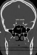
CAPT Butler believes the blood in the mask is key, and that this is barotrauma of the sphenoid sinus, probably complicated by mask squeeze. His rationale is that the sphenoid sinus is immediately adjacent to the optic chiasm and there are other case reports of peripheral neuritis related to barotrauma, though none like this. He sent me an image, which is attached. The sphenoid sinus is labeled SpS, and the optic canals are noted.

None of the practitioners I consulted with had heard of a similar case.

Recommendations:
Dr. Freiberger: follow up with an ophthalmologist; already completed.

Dr. Moon: Follow up with a neurologist to rule out non-diving-related neurological causes.

CAPT Butler: See if the ophthalmologist or another practitioner will order a CT scan of the sinuses. Depending on how far out from the injury the CT is performed, it may show residual blood/fluid in the sphenoid sinus and would probably be diagnostic in that case.

Questions that remain: Aviayu, what day did you make the dive, and how long after the dive did you see the ophthalmologist? Can you verify again that you have had no acute or chronic sinus problems, perhaps allergies, a cold, or infection? Are you a smoker? Finally, if it's been a short enough time since the dive (less than a week or so), would it be possible for you to get a CT scan of your sinuses?

This is a fascinating case. Aviayu, I think we'd all like to see this through to a conclusion and I know all the practitioners here will help in any way possible.
 

Attachments

  • Sphenoid2.jpg
    Sphenoid2.jpg
    23.5 KB · Views: 78
DocV, yes it's my understanding that direct ophthalmoscope examination cannot exclude having a detachment. If suspicions persist despite a clean regular exam, IMO, a US may be useful. In this case, mention of a "curtain" is suspicious although that must be weighed against the absence of "sparkles" which is also commonly reported.

-----------

I concur with CPT Butler* because it aligns with my experiences with panda/raccoon eyes in trauma cases. Effective ear/sinus equalization does appear to be a concern because this diver has admitted that nose pinching doesn't really work for him so that he only swallows. If possible inadequate sinus equalization is tolerated, then this raises questions of tolerance for inadequate mask equalization.

The obvious concern is for the optic nerves being affected by mechanical pressure; onset and resolution of blindness were related to depth. In addition to detecting frank blood as mentioned by CPT Butler, a CT may also reveal whether there is reason to suspect a further anatomical contribution to make the case so unusually severe; i.e. an irregular structure which may affect nerve passage or blood supply.


*I've seen his online open copy of a 2004 AlertDiver article on breathholding.

.
 
DocV, yes it's my understanding that direct ophthalmoscope examination cannot exclude having a detachment.

Not the question I asked you, cutlass. The question I posed was "a special ultrasound examination" (high-resolution ultrasound imaging?) v ocular fundus photography.

Regards,

DocVikingo
 
Thanks for all the effort, Duke Dive Medicine.

Indeed this is a fascinating, and worrisome, case.

Ditto "we'd all like to see this through to a conclusion and I know all the practitioners here will help in any way possible."

Regards,

DocVikingo
 
Not the question I asked you, cutlass. The question I posed was "a special ultrasound examination" (high-resolution ultrasound imaging?) v ocular fundus photography.

Regards,

DocVikingo

They're complementary. Under your conditions, fundus photography would be preferred. US would also work here but is also especially suited for obscured conditions; e.g. facial trauma.

Common, direct ophthalmoscope inspection may be inadequate because of a limited field of view and its dependence on simply reflected light to detect changes in the red reflex. Indirect and fundus photography overcome these deficiencies.
 
Duke Dive Medicine, I'd like to thank you for your excellent recap AND for the effort you've made to run this case past some of the best people in the world to evaluate it.

To me, this is an absolutely fascinating case -- CT results would be very interesting, but it may well be that, as the problem has resolved and there IS a presumptive diagnosis, the OP's physicians (given where he is) may not be interested in pursuing the matter to that point.
 
https://www.shearwater.com/products/peregrine/

Back
Top Bottom