

gert7to3
Contributor
- Messages
- 1,169
- Reaction score
- 121
- # of dives
- 200 - 499
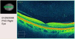
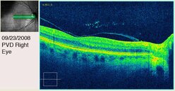
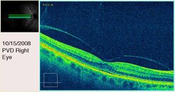
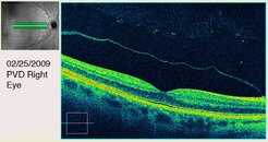
Attached are three OCT images of the PVD progression in my right eye over a period of nine months. The fourth image was taken in February.
The orange band is my retinal pigment epithelium. The yellow-green bands are the layers of my retina. The little depression in the center of the green banded area is my fovea. The fainter green band in the black space above my retina is the vitreous humor detaching from the inner limiting membrane. The second image shows some floaters. The third image was made just before the detachment ended. You can even see that it was pulling my fovea up, which caused a clear distortion in my central vision.
These images were made with a Zeiss Cirris OCT.